
Electrode Geometry
Contents
Electrode Geometry
Extracellular
Orientation and Polarity
Remember that voltage is measured as the difference between two electrodes. Electrodes have a + and - designation. The + is compared to the -. Therefore, the polarity of the voltage measurement across those electrodes will depend on which electrode (+ or -) is in an area of higher voltage relative to the other (Figure 1).
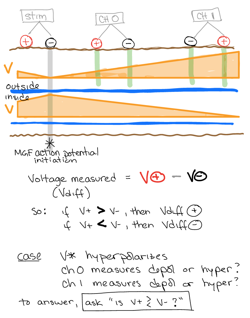
Figure 1 Effects of electrode orientation on polarity of action potential in measurement.
Tracking voltage activity across time and space
The earthworm nervous system provides an excellent way to track the effects of space and time on action potential propogation. By changing the orientation and/or separation between measurement electrodes, you can infer some interesting features of the action potential. The most stunning feature that you can infer is the “width” (in space and time) of the action potential as it travels along the axon (Figure 2).
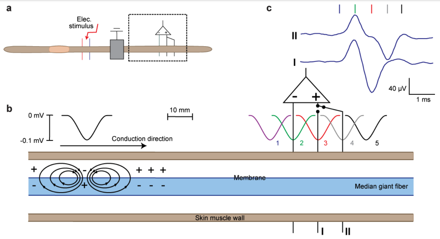
Figure 2 From Kladt et al: Spatial dimensions of action potentials. (a) Experimental setup. (b) The conduction of action potentials. An electrical stimulus leads to the generation of an action potential by depolarizing the membrane; in this case the median giant fiber. This depolarization leads to a negative potential wave [outside the membrane] caused by current loops. As the negative potential wave is conducted along the fiber, the amplitude of the recording depends on the difference between the two electrodes (+/-). This difference is dependent on electrode distance, i.e., if the negative potential wave fits in between the electrodes (setting II), there is no potential difference measured for a short period of time and this results in a plateau showing up in the recordings. (ie. The plateau is the visible result of the negative potential wave fitting in between the two electrodes)(c) Examples of evoked CAPs with electrodes placed at distances of 1 and 2 cm (I/II).
This experiment is a powerful and simple demonstration that experimental design has to be thoughtful and that the results have to be questioned thoroughly. Even the worm action potentials are big in terms of spatial wave lengths (1-1.5 cm). In humans, these can even be bigger, e.g., 10cm for fibers with 100 m/s conduction velocity and APs of 1ms duration.
This virtual lab website from McGill has another graphic showing this effect
Review of Passive membrane properties
Relevant review for why there is a voltage gradient to measure.
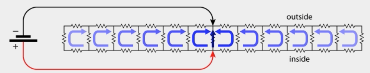
Figure 3 Diagram of current flow (copywrite Bob Wyttenbach)