Week 12. Human Motor Neuron Coding
Contents
Week 12. Human Motor Neuron Coding
In vertebrates, every voltage spike in a motor neuron generates a voltage spike in the muscle fibers it innervates. This is in contrast to the superficial flexor muscle of crayfish, in which graded post-synaptic potentials summed without eliciting an action potential.
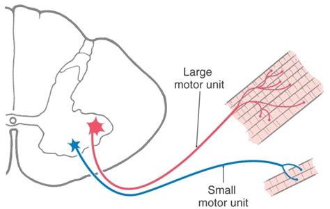
A single motor neuron and the muscle fibers it innervates are called a motor unit. This makes it the smallest functional unit of the nervous system as well as the final output of motor commands. In humans, the only single-unit neural activity that we can measure non-invasively is from motor neurons (albeit indirectly via the muscle fiber spike). A collection of motor units a known as a motor pool. The number of muscle fibers controlled by a single motor neuron varies greatly. Small motor units are those in which a single motor neuron action potential
Today you will be recording the activity of individual neurons in the motor system of yourself and/or each other. Specifically, you will be using differential electrodes on the surface of the skin to pick up the extracellular voltage signal associated with muscle cell action potentials (ie an electromyogram, EMG).
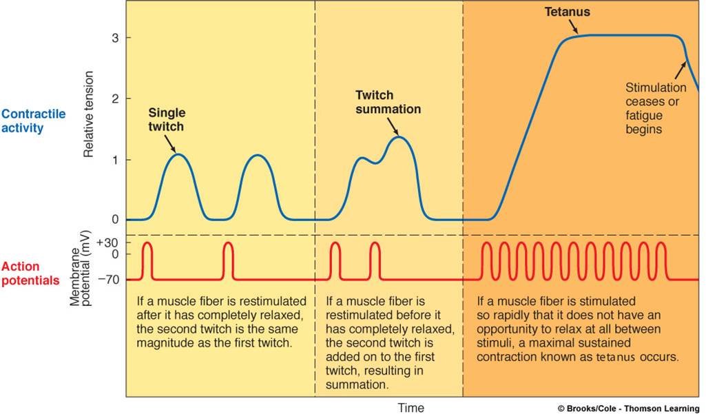
Every time a pre-synaptic motor neuron fires an action potential, the muscle cells that it innervates fire an action potential. This one-to-one relationship enables the muscle action potential to serve as a proxy for the motor neuron action potential. All muscle fibers within a motor unit will generate an action potential at the same time and their amplitude will be summed. Therefore, larger motor units will have larger waveforms in an EMG signal.
Additionally, muscle contraction strength (amount) depends on the rate of muscle action potentials. Temporal summation of action potentials within a single muscle fiber increases the muscle contraction.